


Osteochondrosis is a chronic degenerative-dystrophic disease that develops under the influence of many very different factors. Initially, pathological changes appear in the pulpal nucleus (the internal contents of the intervertebral disc), and subsequently spread to the fibrous ring (the outer shell of the disc) and other elements of the segment of movement of the spine (SDS). This can be a consequence of the body's natural aging process or it can occur against the background of injuries, increased loads on the spine and other causes. In any case, osteochondrosis is only the first stage of destruction of the intervertebral disc and if left untreated, protrusions and hernias form, which often require surgical removal.
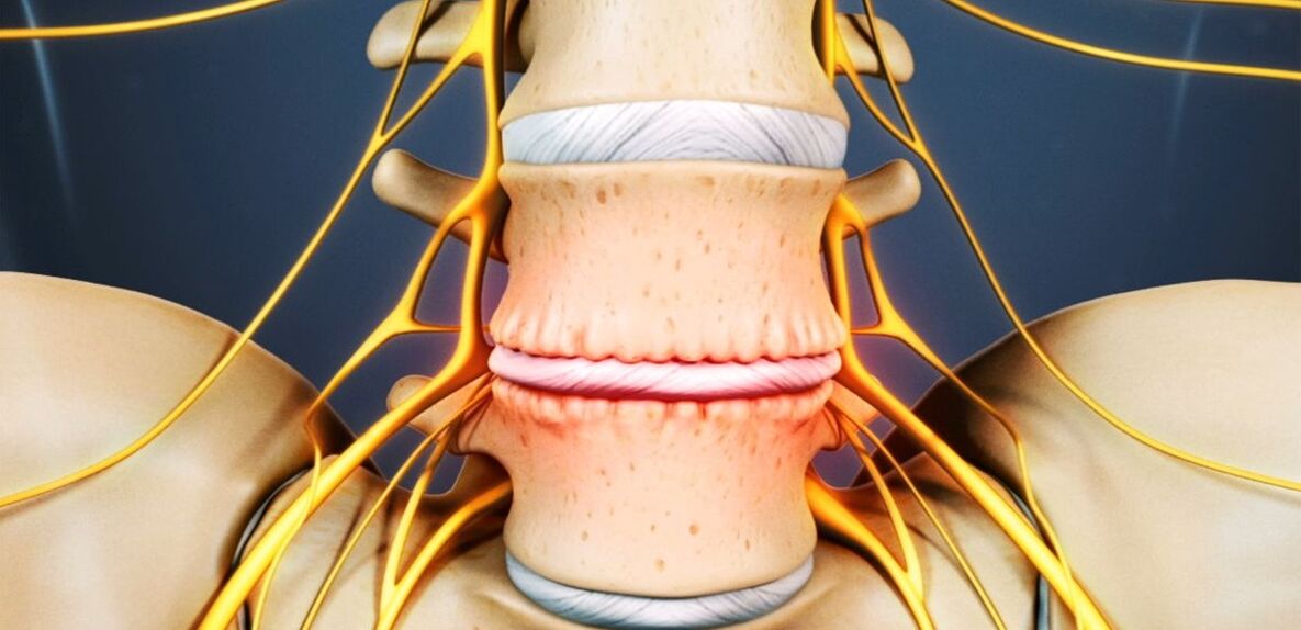
The intervertebral disc is a cartilaginous formation that divides the bodies of the vertebrae and acts as a shock absorber.
Osteochondrosis of the lumbar region: what is it
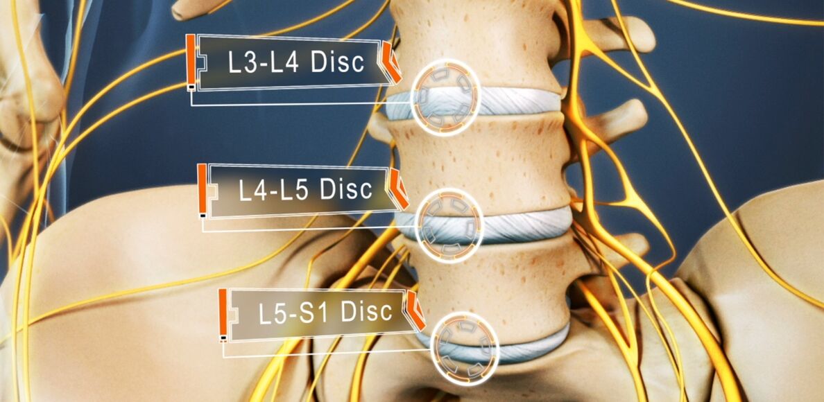
Osteochondrosis affects 48 to 52% of people. And osteochondrosis of the lumbar spine is the most common. The disease can affect any of the intervertebral discs of the lumbosacral spine, several or even all of them. Disks L5-S1, L4-L5 suffer most often, less often L3-L4. The upper lumbar discs (L3-L2 and L2-L1) are much less commonly affected.
The prevalence of lumbar osteochondrosis is due to the fact that the greatest load during the performance of any physical work, especially lifting and carrying weights, walking, running, sitting, falls on the lower back. The lumbar spine consists of 5 vertebrae, which are much larger than the thoracic and cervical vertebrae. Accordingly, the intervertebral discs that separate them are larger in size. The lumbar region usually has a slight anterior curvature (physiological lordosis). This is the last mobile part of the spine and is adjacent to the fixed sacrum, so lumbosacral osteochondrosis is most commonly referred to.
If earlier osteochondrosis was considered an age-related disease, today its first manifestations can be observed at the age of 15-19. Among 30-year-olds already 1, 1% of people suffer from severe symptoms of degenerative-dystrophic changes in the intervertebral discs. And in representatives of the older age group (from 59 years) the clinical manifestations of the disease are already present in 82, 5%. At the same time, the incidence of pathology continues to grow steadily, which is largely due not only to the increase in the average age of the country's population, but also to lifestyle changes that are not for the better.
Reasons for development
Today there is still no consensus on the etiology of degenerative diseases of the spine. However, the basic theory of their development is involutive. According to her, osteochondrosis is a consequence of previous damage to the intervertebral disc and bone structures of the spine, as well as the appearance of inflammatory and other processes. The theory suggests that degenerative changes are genetically predisposed and are in fact inevitable. And their clinical manifestation, especially in young and middle-aged people, is due to the influence of various endogenous and exogenous factors.
Thus, the development of osteochondrosis of the lumbar spine is facilitated by:
- heavy physical labor, especially weight lifting;
- sedentary, sedentary lifestyle;
- any back injuries, including bruises;
- Overweight;
- metabolic disorders;
- posture disorders, deformity of the spine;
- flat feet and other pathologies of the legs;
- pregnancy, especially multiple pregnancy.

Pathogenesis
Regardless of the causes, degeneration of the intervertebral disc occurs when the intensity of the processes of catabolism (cleavage and oxidation of molecules) of matrix proteins begins to exceed the rate of their formation. One of the key points in this process is the malnutrition of the intervertebral discs.
Because, like most cartilages in an adult, they do not have a direct blood supply because they are deprived of blood vessels, the supply of nutrients to them and the removal of metabolic products is by diffusion with sequential compression and relaxation of the disc during movement. The main structure that provides power to the disk are the end plates located on its upper and lower surface.
The end plates themselves are a bilayer formed by cells of cartilage and bone tissue. Accordingly, the cartilaginous side they are adjacent to the disc, and the bone - to the vertebral bodies. They are characterized by good enough permeability, which ensures the exchange of substances between the cells, the intercellular substance of the disc and the blood vessels passing into the vertebral bodies. Over the years, especially with the negative impact of external and internal factors, the structure of the end plates changes and their blood supply decreases, which leads to a decrease in the intensity of metabolism in the intervertebral disc. As a result, its ability to produce a new matrix is reduced, which leads to a progressive decrease in its density with age.
At the molecular level, this is accompanied by:
- reducing the rate of diffusion of nutrients and metabolic products;
- reducing cell viability;
- accumulation of cell breakdown products and altered matrix molecules;
- reduction of the production of proteoglycans (macromolecular compounds responsible for the formation of new cartilage cells and which are the main sources of synthesis of chondroitin sulfates);
- damage to the collagen scaffold.
Possible consequences
As a result of the ongoing changes, the intervertebral disc becomes dehydrated and the pulpal nucleus loses its ability to adequately distribute the loads that fall on it. As a result, the pressure inside the disc becomes uneven and therefore the fibrous ring experiences overload and compression in several places. As this happens with every movement of a person, the annular ring is regularly subjected to mechanical pressure. This leads to adverse changes in it.
Also, often the reduction in the height and elasticity of the disc leads to compensatory changes in the adjacent vertebral bodies. Bone growths called osteophytes form on their surface. They tend to increase in size over time and even merge with each other, excluding the possibility of movement in the affected PDS.
Due to the fact that malnutrition provokes damage to the collagen skeleton, under the influence of the pressure of the pulpal nucleus at certain points, the normal structure of the fibers forming the fibrous ring is disrupted. In the absence of intervention, this eventually leads to cracks and fractures in them. Gradually more and more fibers of the fibrous ring at the site of application of pressure are torn, which leads to its protrusion. This is especially facilitated by the increased loads on the spine. And because the lumbar region takes on the main load during movement and any physical activity, it suffers most often.
The protrusion of the intervertebral disc without permanent rupture of the fibrous ring and with a base size larger than the protruding part is called protrusion. With its complete rupture in one place or another, an intervertebral hernia is diagnosed.
With the destruction of part of the fibers of the fibrous ring, the pressure in the disk gradually decreases, which leads to a decrease in tension and the fibers themselves. This leads to a violation of its fixation and, as a result, pathological mobility of the affected segment for movement of the spine.
The spinal motor segment (SMS) is a structural and functional unit of the spine formed by the intervertebral disc, the adjacent vertebral bodies, their facet joints, ligaments and muscles attached to these bone structures.
Normal functioning of the spine is possible only with proper functioning of the PDS.
Symptoms of osteochondrosis of the lumbar spine
The disease may be asymptomatic for a long time, and then begin to manifest itself as a slight discomfort in the lumbar region, gradually gaining strength. But in some cases, lumbar osteochondrosis begins acutely, immediately provoking severe pain. In most cases, the signs of pathology appear for the first time after 35 years.
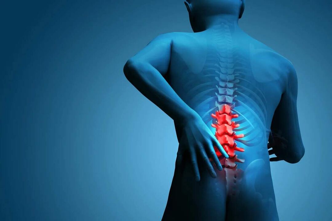
Back pain is the main symptom of the disease. It can be different in nature and can be painful and dull, as well as acute, constant or episodic. But the pathology, especially in the early stages of development, is characterized by alternating periods of exacerbation and remission, and both hypothermia and lifting a heavy object, or unsuccessful, sudden movement can provoke another deterioration in well-being.
The pain is often accompanied by a tingling sensation and tension in the back muscles. They worsen with exercise, sudden movements, lifting weights, bending over and even coughing and sneezing.
If, due to the instability of the vertebral bodies, the root of the nerve extending from the spinal cord is captured by one or another anatomical structure, this will lead to the development of appropriate neurological disorders. Their main manifestations are:
- shooting, severe pain radiating to the sacrum, buttocks, lower limbs or perineum;
- sensitivity disorders of varying severity;
- mobility restrictions, lameness;
- weakness in the muscles innervated by the pinched nerve.
In the lumbar spine, the spinal cord ends at the level of 1-2 vertebrae and passes into the so-called cauda equina, formed by the accumulation of spinal roots. In addition, each of them is responsible not only for the innervation of the muscles, but also for specific organs of the pelvis, so prolonged compression can cause disturbances in the work of the organ. This can lead to the development of impotence, infertility, gynecological diseases, hemorrhoids and other disorders.
The clinical picture of osteochondrosis of the lumbar spine, especially with a long course and the appearance of compression of the spinal roots, largely depends on the level of the lesion, ie which particular disc has undergone degenerative-dystrophic changes.
- Defeat of the disc L3-L4 - pain is given in the anterior-inner parts of the thigh, lower leg and inner ankle. This is accompanied by a decrease in the sensitivity of the anterior surface of the thigh, a decrease in weight or loss of the knee, as well as a decrease in the strength of the quadriceps muscle.
- Defeat of the disc L4-L5 - the pain is given from the upper part of the buttocks to the outer parts of the thigh and lower leg. Less commonly, this is accompanied by the spread of pain in the back of the feet, including 1-3 toes. Decreased sensitivity and muscle weakness are observed in these areas. Sometimes malnutrition and incomplete extension of the big toe develop.
- Disc damage L5-S1 - the pain starts in the middle area of the buttocks and goes down to the heel on the back or back of the thigh and lower leg and can catch the outer edge of the foot, like 4-5 toes. In these areas of the lower extremities there is a decrease in sensitivity, and the gastrocnemius and large gluteus often decrease in size, which is accompanied by their weakness. If the spinal root passing at the level of this PDS is affected, a decrease or loss of Achilles and plantar reflexes may be observed.
Disks L1-L2 and L2-L3 are rarely affected.
The pain that accompanies the disease limits the person and significantly reduces his quality of life. Because they last a long time and recur regularly, if not constantly, this can not but affect the psycho-emotional state. As a result, more than half of patients show signs of chronic emotional stress, depressive disorders and more.
Diagnosis
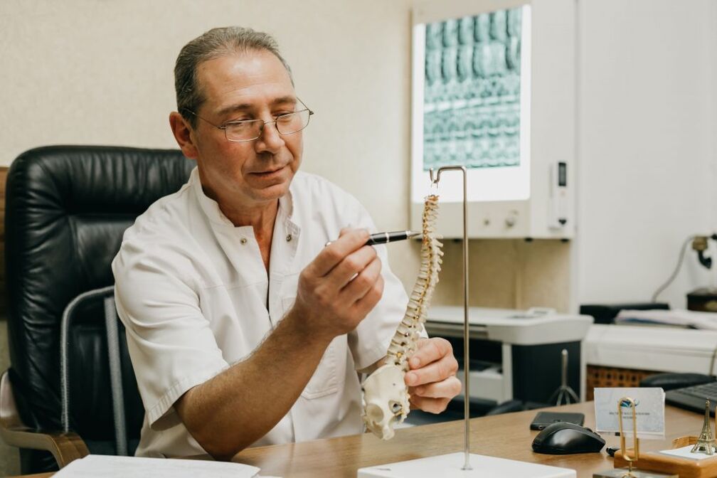
If there are signs of osteochondrosis of the lumbar spine, you should contact a neurologist or vertebrologist. First of all, the doctor collects the anamnesis, which consists in clarifying the nature of the complaints, the characteristics of the pain, the conditions for their occurrence and reduction, the characteristics of the working life of the person and others.
The second stage of diagnosis, performed as part of the first consultation with a doctor, is a physical examination. During it, the doctor assesses the condition of the skin, posture, the depth of the physiological curves of the spine, the presence of its curvature and more. The condition of the muscles around the spine, called paravertebral, must be assessed because they are often painful and over-tense, which is a reflex reaction of the body to inflammation and discogenic pain.
Already on the basis of the data obtained during the examination and questioning of the patient, the neurologist may suspect the presence of osteochondrosis of the lumbar spine. However, in order to exclude possible concomitant pathologies, as well as to confirm the diagnosis and determine the exact level of damage, the severity of degenerative-dystrophic changes in the intervertebral disc and the involvement of bone structures, laboratory and instrumental diagnostic methods are needed.
Laboratory diagnostics
Different types of tests are not crucial in diagnosing osteochondrosis of the lumbar spine. They are more focused on assessing the extent of the inflammatory process and detecting concomitant disorders.
In this way they can be appointed:
- UAC;
- OAM;
- blood sugar test;
- blood chemistry.
Instrumental diagnostics
It has been shown that all patients with suspected lumbar osteochondrosis have:
- X-ray of the lumbar spine in two projections - allows you to determine the structure of bone structures, to detect abnormalities, formed osteophytes, changes in the facet joints and more. ;
- CT - allows you to detect changes in bone structures at earlier stages of development from X-rays, as well as to identify indirect signs of osteochondrosis;
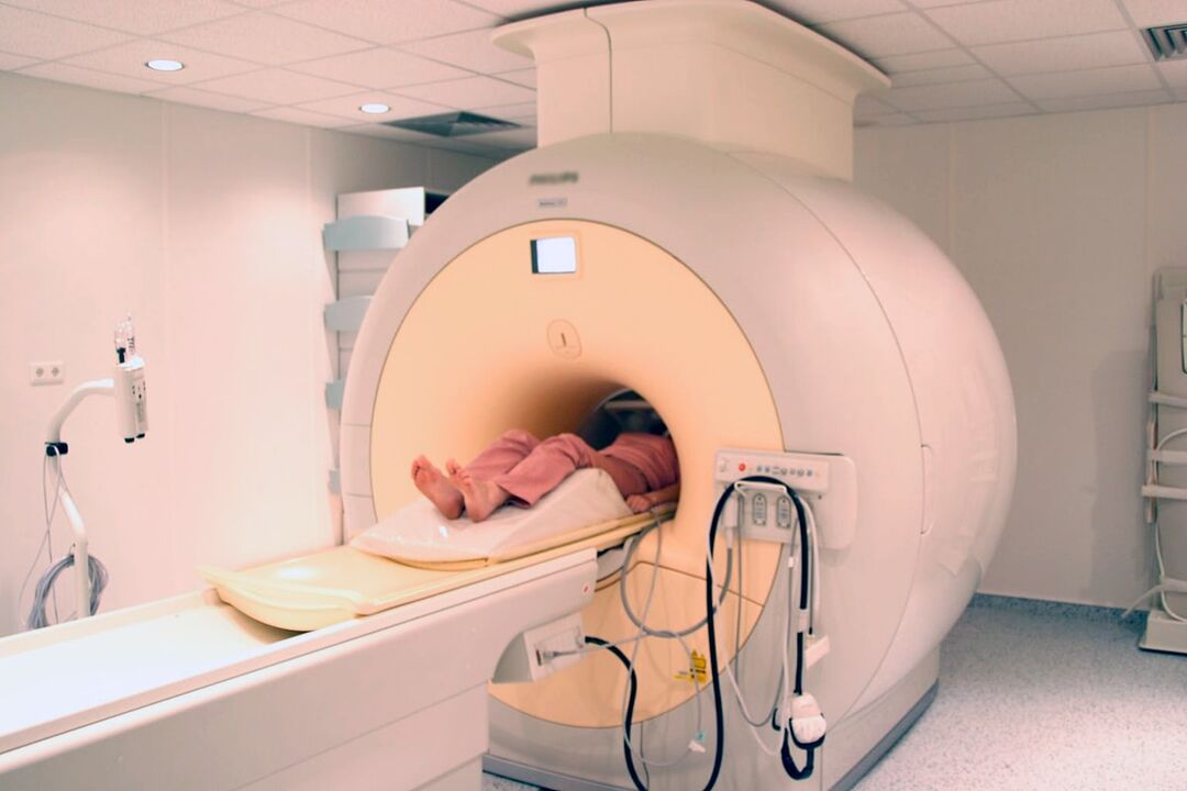
- MRI is the best method for diagnosing pathological changes in cartilage formations and other soft tissue structures, which makes it possible to detect even the smallest changes in the intervertebral discs, ligaments, blood vessels and spinal cord and to accurately assess their severity and potentialrisks.
In addition, it may be recommended:
- densitometry - a method for determining bone density, which makes it possible to diagnose osteoporosis, which is especially common in the elderly;
- myelography - allows you to assess the condition of the CSF pathways of the spinal cord and the degree of damage to the protruding disc, which is especially important in the presence of an already formed intervertebral hernia of the lumbar spine.
Treatment of lumbar osteochondrosis
When osteochondrosis is diagnosed, as a rule, all patients are initially prescribed conservative therapy, provided that there is no pronounced and progressive neurological deficit. But her character is chosen strictly individually.
Because the disease is chronic and the regenerative capacity of the intervertebral discs is extremely limited, especially in severe degenerative-dystrophic changes, the main goals of therapy are to stop their further progression and eliminate the symptoms that disturb the patient. Therefore, treatment is always complex and includes:
- drug therapy;
- manual therapy;
- physiotherapy;
- exercise therapy.
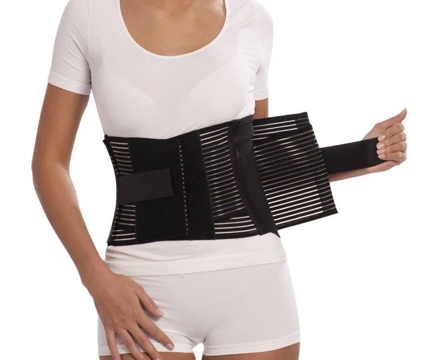
In the acute period, patients have been shown to limit physical activity or even adhere to bed rest for 1-2 days. This will help relax the muscles and reduce the pressure inside the disc. If you have to sit, walk or do physical work for a long time, you should wear a stabilizing lumbar corset.
After the end of the acute period and during remission of the disease, on the contrary, it is important to move as much as possible, but with caution and excluding the increased load on the lower back. Patients will need to acquire skills for proper sitting, lifting objects from the floor, carrying heavy loads, as all this affects the course of the pathology. It is important to avoid tilting and sudden movements, lifting something off the floor or low surfaces after bending your knees, not bending over. You should only sit with a straight back of a chair that supports your back well. In addition, during sedentary work, it is important to take regular breaks for a short workout. It is important to avoid falls, jumps, jogging and hypothermia.
In osteochondrosis it is important to keep the body weight in optimal limits, and in obesity is shown diet and exercise appropriate for the patient's condition, as being overweight creates an increased load on the lower back and causes faster progression of pathological changes. in the discs.
On average, conservative therapy is usually prescribed for 1-3 months, although it can last longer. But even after completing the main course prescribed by your doctor, you will need to continue taking a number of medications, exercise therapy, and follow lifestyle recommendations.
Medical therapy
The main components of drug therapy are individually selected drugs from the group of NSAIDs. In choosing them, the doctor takes into account not only the severity of the pain syndrome and the course of the inflammatory process, but also the nature of concomitant diseases, especially the digestive tract, as NSAIDs with prolonged use can adversely affect the condition of their mucous membranes and provoke exacerbation. various pathologies of the digestive system.
It is necessary to use NSAIDs in acute low back pain and immediately after their appearance. Preferably after 1-2 days. Depending on the severity of the patient's condition, they can be administered intramuscularly, in the form of rectal suppositories, topical agents and oral forms. The duration of administration should not exceed 2 weeks. In the future, an individually selected drug is taken on request, but tries to avoid frequent use.
Recently, drugs as active ingredients that include selective cyclooxygenase-2 inhibitors have become increasingly preferred.
Patients are also prescribed drugs from the following groups:
- muscle relaxants - help to relax overly tense muscles and thus reduce back pain;
- chondroprotectors - improve the course of metabolic processes in the intervertebral disc (especially effective when started in the earliest stages of lumbar osteochondrosis);
- B vitamins - contribute to improving nerve conduction;
- antidepressants and anxiolytics - used in prolonged osteochondrosis, leading to depression, chronic fatigue and other psychological disorders.
In case of very severe pain, especially of neurological origin, therapeutic blockades are performed. These include the administration of anesthetics in combination with corticosteroids at points near the pinched nerve, leading to rapid elimination of pain. However, the procedure can only be performed in a medical facility by specially trained healthcare professionals, as it involves a risk of complications.
Manual therapy
Manual therapy allows not only to improve the quality of blood circulation in the affected area, but also to significantly reduce the severity and duration of pain in osteochondrosis. It effectively relieves muscle tension and allows you to remove functional blocks, which significantly increases mobility in the affected SMS.
Also, through well-conducted manual therapy, it is possible not only to increase the distance between the vertebrae, to return them to their anatomically correct position, but also to release the compressed nerve roots. As a result, the pain is quickly eliminated and the neurological disorders disappear. It also reduces the likelihood of complications and disorders of the internal organs.
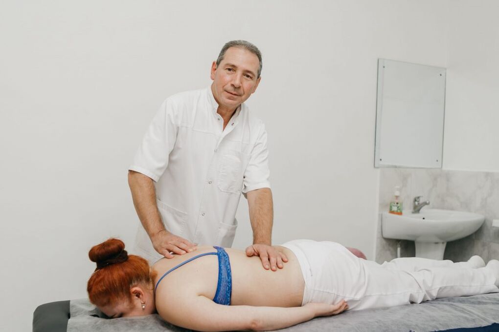
Additional positive properties of manual therapy are improving mood, strengthening immunity, activating the body's natural recovery mechanisms and increasing efficiency. Usually after the 1st session there is a noticeable improvement in well-being, and in the future the effect becomes more pronounced. As a rule, the course consists of 8-15 sessions and it is important to complete it by the end, even with complete normalization of well-being.
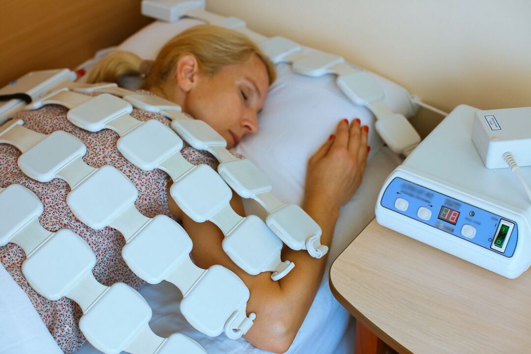
Physiotherapy
After the disappearance of the acute inflammation, courses of physiotherapeutic procedures are shown, which not only help to reduce the pain, but also improve the microcirculation, nutrition and the course of the reparative processes in the field of degenerative-dystrophic changes. Most often patients are prescribed:
- drug electrophoresis;
- electrical neuromyostimulation;
- ultrasound therapy;
- laser therapy;
- magnetic therapy;
- UHF.
What specific methods of physiotherapy will give the best effect, the frequency of their application, the duration of the course and the possibility of combining with other types of exposure is determined individually for each patient.
Traction therapy gives very good results in osteochondrosis of the lumbar spine. Thanks to it, it is possible to increase the distance between the bodies of the vertebrae, which instantly reduces the load on the affected discs. After the session, the patient should wear an orthopedic corset to consolidate the results.
exercise therapy
After eliminating the acute pain, the treatment program must be supplemented with therapeutic exercise. Its main tasks are stretching the spine and relaxing the spasmodic muscles of the lower back. Also, therapeutic exercises help strengthen the muscular corset, create a reliable support for the spine and improve posture. In the course of this, blood circulation is inevitably activated and metabolic processes are improved, which has a beneficial effect on the nutrition of the discs.
A set of exercises is selected for each patient individually in accordance with the degree of degenerative-dystrophic changes, the level of physical development of the patient, the nature of the accompanying disorders, age and other factors. Initially, it is recommended to study under the guidance of an experienced exercise instructor.
All patients with degenerative changes in the spine are recommended to visit the pool 2-3 times a week, as swimming lessons minimize the load on the spine, but allow you to effectively strengthen the muscles of the back.
Thus, osteochondrosis of the lumbar spine is one of the most common diseases. At the same time, it is able to deprive a person of the ability to work for a long time and even cause damage due to the development of complications. That is why it is important not to ignore the first symptoms of the disease when it is easiest to deal with it. With the onset of pain and even more tingling, limited mobility, back pain, you should contact a neurologist as soon as possible, undergo the necessary examination and begin treatment. In this case, it will be possible to stop the pathological process and return to a normal, full life without pain and significant limitations.